Spatial Transcriptomics: Mapping Disease at the Cellular Level
Introduction
Understanding disease requires more than identifying which genes are active in a tissue it also requires understanding where those genes are expressed within the biological architecture of the tissue itself. The spatial arrangement of cells within organs and tissues plays a critical role in regulating biological processes such as cell signaling, immune responses, and tissue development. Disruption of this spatial organization can contribute to the development and progression of many diseases, including cancer, neurodegenerative disorders, and inflammatory conditions.
Traditional genomic methods such as bulk RNA sequencing and even single-cell RNA sequencing have provided powerful insights into gene expression patterns. However, these methods often require dissociation of tissues into individual cells, which removes information about the spatial relationships between cells and their surrounding microenvironment. Without spatial context, it becomes difficult to fully understand how cellular interactions influence disease processes.
Spatial transcriptomics is an emerging technology that addresses this limitation by enabling the measurement of gene expression within intact tissue sections while preserving information about the physical location of cells. By integrating molecular profiling with spatial information, spatial transcriptomics allows researchers to generate detailed maps of gene activity within tissues.
This approach is increasingly being applied across multiple biomedical fields, including oncology, neuroscience, and immunology. As these technologies mature, spatial transcriptomics may also contribute to the development of precision pathology, where molecular and spatial information are integrated to improve disease diagnosis and treatment planning.
What Spatial Transcriptomics Is
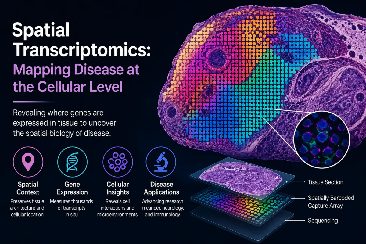
Spatial transcriptomics refers to a group of molecular technologies that measure RNA expression while retaining information about the spatial location of transcripts within a tissue sample. Unlike conventional RNA sequencing approaches that analyze homogenized tissue samples, spatial transcriptomics preserves the architecture of the tissue during analysis.
In practical terms, this means that researchers can determine not only which genes are expressed but also where within the tissue those genes are active. This spatial information is crucial because the biological function of cells is strongly influenced by their position relative to neighboring cells, blood vessels, and structural components of the tissue.
Spatial transcriptomics techniques combine several key components:
Histological imaging of tissue sections
Spatially barcoded capture probes or imaging-based detection methods
RNA sequencing technologies capable of measuring thousands of transcripts simultaneously
One widely used approach involves placing a thin tissue section onto a slide containing an array of spatially barcoded probes that capture messenger RNA (mRNA) molecules. When the captured RNA is sequenced, the barcode associated with each transcript allows researchers to determine the original spatial position of that gene expression signal within the tissue.
Advances in spatial transcriptomics technologies have improved both the resolution and the number of detectable genes. For example, high-definition spatial transcriptomics methods can capture RNA molecules from tissue sections using dense arrays of spatially barcoded beads, enabling high-resolution mapping of gene expression within complex tissues.
These approaches allow researchers to reconstruct spatial maps of gene activity across entire tissue sections, revealing previously unrecognized cellular interactions and functional regions.
Why Tissue Architecture Matters in Disease
The importance of tissue architecture in disease has long been recognized in pathology. Traditional histopathology relies on microscopic examination of tissue morphology to identify structural abnormalities that indicate disease.
However, morphology alone does not fully capture the molecular mechanisms underlying disease processes. Many pathological changes are driven by alterations in gene expression that occur within specific cell populations or microenvironments.
The spatial organization of cells influences several critical biological processes:
Cell-cell communication - Cells interact with neighboring cells through signaling molecules and receptor-mediated pathways.
Microenvironmental regulation - Local gradients of oxygen, nutrients, and immune signals influence gene expression.
Functional compartmentalization - Many tissues contain specialized regions with distinct cellular functions.
For example, in tumors, the spatial arrangement of cancer cells, stromal cells, and immune cells forms a complex tumor microenvironment that strongly influences disease progression and therapeutic response.
Traditional transcriptomic methods cannot fully capture these interactions because they lack spatial resolution. Spatial transcriptomics allows researchers to investigate how gene expression patterns are organized across tissue structures and how these patterns change in disease.
Applications of Spatial Transcriptomics
Spatial transcriptomics technologies are being applied in a growing number of biomedical research fields. By providing spatially resolved molecular data, these methods offer new insights into disease biology and tissue organization.
Oncology
One of the most prominent applications of spatial transcriptomics is in cancer research. Tumors are highly heterogeneous structures composed of multiple cell types, including cancer cells, immune cells, fibroblasts, and endothelial cells. The spatial distribution of these cells plays an important role in tumor progression and treatment response.
Spatial transcriptomics allows researchers to map gene expression across tumor tissues and identify distinct cellular niches within the tumor microenvironment. These analyses can reveal:
Regions of tumor proliferation
Immune cell infiltration patterns
Stromal cell interactions
Hypoxic microenvironments
Such insights can help researchers understand how tumors evade immune surveillance or develop resistance to therapy. Spatial transcriptomic studies have also identified molecular gradients within tumors that may influence metastatic potential.
In addition, spatial mapping of gene expression can support the development of biomarkers that reflect the spatial organization of tumors, potentially improving patient stratification for targeted therapies or immunotherapy.
Neuroscience
The human brain contains an extraordinary diversity of cell types organized into complex anatomical structures. Understanding the spatial distribution of gene expression within these structures is essential for studying brain development, neural circuitry, and neurological disease.
Spatial transcriptomics has been used to generate detailed molecular maps of the brain, identifying distinct neuronal populations and their spatial relationships. These studies have contributed to efforts to build comprehensive atlases of the human brain.
In neurodegenerative diseases such as Alzheimer’s disease and Parkinson’s disease, spatial transcriptomics can reveal how pathological changes spread across brain regions. By mapping gene expression changes in affected tissues, researchers can identify cell populations involved in disease progression and uncover potential therapeutic targets.
Spatial transcriptomic approaches have also been applied to investigate the cellular responses to brain injury and inflammation, providing insights into the interactions between neurons, glial cells, and immune cells.
Immunology
Immune responses are highly dependent on spatial organization within tissues. Immune cells migrate through tissues, form localized clusters, and interact with other cell types to coordinate responses to infection or injury.
Spatial transcriptomics enables researchers to study these interactions at a molecular level. For example, researchers can map the locations of specific immune cell populations within inflamed tissues and analyze how gene expression varies across different microenvironments.
This capability is particularly valuable for studying chronic inflammatory diseases such as rheumatoid arthritis, inflammatory bowel disease, and autoimmune disorders. By identifying spatial patterns of immune activation, researchers can gain insights into disease mechanisms and identify potential therapeutic targets.
Spatial transcriptomics is also being applied to vaccine research and infectious disease studies, where understanding the spatial dynamics of immune responses can inform vaccine design and immunotherapy strategies.
Future Potential: Toward Precision Pathology
As spatial transcriptomics technologies continue to evolve, they may transform the practice of pathology by integrating molecular data with tissue architecture.
Traditional pathology relies primarily on morphological evaluation of stained tissue sections. While this approach remains essential, it does not capture the full molecular complexity of disease processes.
Spatial transcriptomics offers the possibility of precision pathology, where molecular information about gene expression is layered onto histological images of tissues. In such a framework, clinicians could evaluate both the structural and molecular features of disease within a single tissue sample.
Potential applications of spatial transcriptomics in clinical pathology include:
Improved classification of tumors based on spatial gene expression patterns
Identification of prognostic biomarkers associated with specific cellular niches
Detection of microenvironmental signatures associated with treatment response
Enhanced understanding of disease progression within tissues
As datasets from spatial transcriptomics studies grow, computational tools including machine learning approaches may be used to integrate spatial molecular data with clinical information. This integration could lead to more precise diagnostic categories and better prediction of therapeutic outcomes.
Implementation Challenges
Despite its promise, several challenges remain before spatial transcriptomics can be widely adopted in clinical settings.
Technical Complexity
Spatial transcriptomics experiments require specialized equipment, sequencing technologies, and computational pipelines for data analysis. Interpreting spatial gene expression data often involves complex bioinformatic workflows.
Cost Considerations
Current spatial transcriptomics platforms remain relatively expensive, limiting their accessibility in many research and clinical laboratories.
Data Integration
Combining spatial transcriptomics data with other types of molecular information such as genomic mutations, proteomic data, and clinical records requires robust computational frameworks and standardized data formats.
Clinical Validation
Before spatial transcriptomics can be integrated into routine clinical practice, studies must demonstrate its clinical utility in improving diagnostic accuracy or guiding treatment decisions.
Addressing these challenges will require collaboration among researchers, clinicians, technology developers, and healthcare systems.
Future Research Directions
Several areas of research are likely to shape the future development of spatial transcriptomics.
Higher-resolution technologies capable of achieving true single-cell or subcellular spatial mapping of gene expression.
Integration with other spatial omics technologies, such as spatial proteomics and spatial metabolomics, enabling multi-omics analysis within intact tissues.
AI-driven analysis tools that can interpret large spatial datasets and identify clinically relevant patterns.
Clinical translation studies that evaluate how spatial transcriptomic information can inform treatment decisions or disease prognosis.
As these advances occur, spatial transcriptomics may become a foundational technology in the broader field of spatial biology.
Conclusion
Spatial transcriptomics represents a significant advancement in molecular biology by enabling researchers to map gene expression within the structural context of tissues. By preserving spatial information, these technologies provide new insights into cellular organization, tissue function, and disease mechanisms.
Applications in oncology, neuroscience, and immunology have already demonstrated the potential of spatial transcriptomics to uncover complex cellular interactions that were previously difficult to observe. As technologies continue to improve, spatial transcriptomics may also contribute to the development of precision pathology, where molecular and spatial information are integrated to guide diagnosis and treatment.
While technical and clinical challenges remain, ongoing research is rapidly expanding the capabilities of spatial transcriptomics. For clinicians, researchers, and healthcare leaders engaged in precision medicine, understanding this emerging technology will be essential for navigating the next generation of molecular diagnostics and disease mapping.
References
Vickovic S, et al. High-definition spatial transcriptomics for in situ tissue profiling. Nature Methods. 2019.
Marx V. Method of the Year: spatially resolved transcriptomics. Nature Methods. 2021.
Lim HJ et al. Spatial transcriptomics technologies and applications. BMC Genomics.
Lein E et al. The promise of spatial transcriptomics in biomedical research. Science.
Rao A et al. Spatial transcriptomics in disease biology. Nature Reviews Genetics.
